Seeing Red: Your Blood Under the Microscope
Not a lot of people could handle seeing their own blood, but there are not a lot of people who had examined their own blood under the compound light microscope either. Blood to humans is just like chlorophyll is to plants, since it is our blood that carries nutrients from the food we eat around our body and takes away what we don’t need. Blood also contains corpuscles which help fight diseases. With all the importance of blood, though, we seldom ever think about it unless we get cut or wounded.Before you begin looking at your blood under a high power microscope objective lens, let’s learn about what you could see in a blood sample first. This will help you understand what you are going to be examining under the blood microscope.
The red corpuscles in your blood carry oxygen to the parts of your body and clean up the waste materials like carbon dioxide. These red cells contain haemoglobin, which gives blood its red color. White blood cells on the other hand protect your body from diseases. Blood also contains substances that trigger homeostasis or blood coagulation. Blood coagulations help stop us bleeding to death in case we are wounded or cut by hardening around the cut or wound and acting like a plug.
Blood examination with the help of a microscope plays a very important role in the identification of diseases. Medical technicians study blood samples of patients to help identify their diseases. An RBC or WBC count would be able to get the actual number of red blood cells or white blood cells in your blood. Once the doctor had identified the sickness, they might take blood samples regularly from their patient to see how they are responding to the medical treatment.
Now that you have learned the basics, let us begin the microscope activity by taking a sample of your blood to look under the microscope. Make sure that the needle you are going to use to prick your finger is clean and sterilized. Nowadays you could buy small lancets that people use with home machines that could take their blood sugar count, so you could use one of those lancets from your local drugstore. However, using a normal pin or needle would be fine as long as you wash it with alcohol and run it through an open flame before using it. Wash your hands before pricking you finger.
Once a sizable amount of blood had welled on your finger, place some of it on a blank microscope slide. Use another slide to smear it onto your first slide. Then place a cover slip over the blood specimen. This will protect the expensive microscope objective lens from coming into contact with the biological blood specimen and ruining the sensitive coatings on the lens.
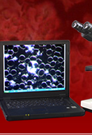
Always begin looking at objects under the microscope using the low power objective first so that you could focus on your sample better as well as find the location to begin viewing. Under the low power objective, you could see that red corpuscles are just faintly red in color. This is because there are five billion red cells in one cubic centimetre of your blood and all those cells are just contributing to the deep red color of your blood. People who have low red cell count usually suffer from anaemia. The doctor would be able to identify what is wrong with an anaemic person by looking at their blood cells under the blood microscope.
White blood cells aren’t as plentiful as red blood cells, counting about one for every five hundred red corpuscle in our blood. Doctors would be able to tell if there is something wrong with their patients if there are too many white corpuscles in their blood samples. It would then be the doctor’s job to track down the part of the body that these white blood cells are targeting to know which part is infected with the disease.
White blood cells are larger than their red counterpart and even contain nuclei. Leucocytes, as white blood cells are called, have different types as based on the shape of their nuclei. If you want to see a polymorphonuclear (having many forms) structure of a leucocyte, you could dilute some pen ink with water and place a drop on the corner of your slide before covering it.
Looking at your own blood under the high power blood microscope may be a fun activity, but it also bears a lot of importance in the field of medicine. Doctors examine blood samples under a high power microscope not only to identify if a person has anaemia by their red blood cell count, but also to be able to pinpoint the center of a disease by keeping track of which body part has more white blood cells than normal. This is just one of the many uses the medical profession has for this type of microscope.


