The Use of the Microscope in Bacteriology
The invention of the microscope opened the door to another world for scientists to pass through to look at organisms or things too small to be seen by the naked eye. The simple microscope was particularly helpful to Anton van Leeuwenhoek in his study about bacteria. Being the first man to observe microorganisms (particularly those living in water) and write down his observations about them, Leeuwenhoek is consequently called the “Father of Microscopy”. It was Leeuwenhoek’s work with the early microscope that set the stage for future development of modern microscopy.Later on, a French chemist called Louis Pasteur discovered microbes that caused fermentation by looking at a sample of milk that had gone sour under the microscope. By studying these microbes and determining where they come from, Pasteur had been able to develop a way of sterilizing liquid so that it wouldn’t ferment. This process is called pasteurization and is named after him. Many of our common grocery store items, such as milk and beer are pasteurized to kill the microscopic bacteria that would shorten their shelf life to a matter of days.
Another significant discovery about microbes was made by a Scotch surgeon named Joseph Lister. Lister had read and admired Pasteur’s research reports and when his patients died from the festering of wounds, Lister began to wonder if the microbes that Pasteur had studied can cause festering as well. Because of Lister’s studies, surgeons began to be vigilant about cleanliness when operating on a patient. The practice of using antiseptics began in this time as well.
But bacteriology or the study of bacteria (a specialization of microbiology) as we know began with a German physician, Robert Koch, who studied harmful bacteria. Starting out with the study of anthrax, Koch had been able to develop ways of observing bacteria. One of his contributions to the field of bacteriology is the way to get only one kind of bacteria to examine under the compound light microscope. These are called pure culture, and Koch found a way to grow his own bacteria on a medium (usually food) to solve this problem. Koch had also laid down rules for bacteriologists to follow before they could attribute any kind of disease on a certain kind of bacteria. First, the sample of bacteria they are studying would have to come from an infected part of an animal suffering from the disease. These bacteria must be grown in pure culture, out of the body of its host. Consequently, healthy animals injected with these cultured bacteria must catch the disease as well.
Aided by Koch’s findings, bacteriologists began examining all sorts of microbes that might be harmful to man, animals and even crops. Taking samples and placing them in Petri dishes to observe under the tissue culture microscope, bacteriologists looked for the causes of certain diseases and set out to find a way to stop these microbes from harming their host further.
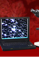
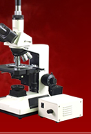
The typical compound light microscope has objectives that face downward and pick up light that is transmitted through the biological specimen on the microscope slide. This is different from the tissue culture microscope that is made specially for viewing cell cultures grown in Petri dishes. This type of microscope has inverted objectives that are under the petri dish and face upward to look into the bottom of the dish. This allows examination of the cell culture from the bottom of the petri dish where the culture first begins to grow. Both use transmitted light microscopy where the light is passed through the specimen. Other methods of viewing bacteria and culture smears may entail the use of the phase contrast microscope. Using phase is often the method of choice when it is desired to not stain the fresh biological specimen.
Some kinds of the bacteria that they had discovered grew very slowly in the body of the host and are stationary in nature. Two of the first bacteria to have been discovered belong to this kind. The first is the bacteria that cause tuberculosis which had been discovered by Koch himself in 1882. Another one is the leprosy bacteria, which grows in the skin.
On the other hand, some bacteria have flagella, or whip-like appendages, that help them move around the body of their host. One of the harmful bacteria with flagella is the cholera vibrio, discovered by Koch in 1884.
In keeping with the saying: know your enemy, the identification of microscopic bacteria that cause diseases had helped scientists develop a means to prevent and cure sickness. The optical microscope has been an indispensable tool in helping scientists in bacteria identification and classification. Using the compound light microscope to aid their studies, scientists had been able to develop medicine and treatment for sickness that had killed off so many people before. The development of the compound microscope as well as the tissue culture (cell culture) microscope has been instrumental in aiding humanity to control diseases caused by bacteria and other microbes.


